Introduction
X-ray technology has come a long way since its invention in 1895. This revolutionary imaging method has been used for over a century to diagnose and treat a variety of medical conditions. But how did it all begin? Who were the pioneers behind this groundbreaking technology? And what impact has it had on modern medicine? This article will explore these questions and more as we take a detailed look at the history and development of X-ray technology.
A Historical Overview of the Invention of X-Ray Technology
The invention of X-ray technology can be traced back to 1895, when German physicist Wilhelm Roentgen discovered that a mysterious type of radiation could pass through solid objects. After experimenting with an evacuated glass tube, he found that the radiation was able to penetrate human skin and produce an image on photographic film. Roentgen named this new form of radiation “X-rays”, and his discovery marked the beginning of the era of modern diagnostic imaging.
In the early years of X-ray technology, scientists and physicians began to explore ways to use this new tool for medical purposes. The first practical application of X-rays was in 1896, when they were used to diagnose broken bones. This quickly led to the widespread adoption of X-ray imaging as a diagnostic tool, and by the early 20th century X-rays were being used to diagnose a variety of medical conditions.
In the modern era, X-ray technology has become an invaluable tool for diagnosis and treatment. Advances in technology have made X-ray imaging faster, safer, and more accurate than ever before. Digital X-ray imaging, for example, produces high-quality images with minimal radiation exposure. In addition, computer-aided detection (CAD) systems can help radiologists to identify abnormalities quickly and accurately.
Examining the Pioneers Behind X-Ray Technology
While Wilhelm Roentgen is often credited as the inventor of X-ray technology, there were many other pioneers who helped to shape the development of this revolutionary imaging method. One of the most influential figures was Marie Curie, who conducted research into the properties of X-rays and their medical applications. Her work provided the foundation for further developments in X-ray imaging, and she is remembered as one of the most important figures in the history of X-ray technology.
Other pioneers of X-ray technology include German physicist Max von Laue, who developed the first X-ray diffraction technique; American physicist Arthur Compton, who studied the scattering of X-rays; and British physicist William Henry Bragg, who developed the first X-ray spectrometer. All of these scientists played an important role in the development of X-ray technology, and their contributions are still being felt today.
Exploring the Impact of X-Rays on Modern Medicine
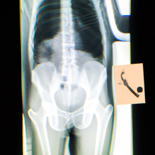
Today, X-ray imaging is an essential part of modern medicine. It is widely used for diagnostic purposes and has become an invaluable tool for the detection and treatment of a wide range of medical conditions. X-rays can be used to detect fractures, tumors, and other abnormalities, as well as to assess the health of internal organs and structures. They can also be used during surgery to help surgeons locate and remove tumors or other abnormalities.
In addition, X-rays are often used to guide minimally invasive treatments, such as biopsies or catheterizations. X-ray imaging can also be used to monitor the progress of certain treatments, such as chemotherapy or radiation therapy. All of these uses demonstrate the powerful impact that X-ray technology has had on modern medicine.
The Science Behind X-Ray Technology
X-rays are a form of electromagnetic radiation, similar to visible light. They have much higher energy levels than visible light and can penetrate solid objects, such as human tissue. X-rays are produced when an electrical current passes through a metal target, such as tungsten or molybdenum. This process causes the metal atoms to emit X-ray photons, which can then be detected by special X-ray films or digital detectors.
There are several different types of X-ray imaging techniques, including conventional radiography, computed tomography (CT), fluoroscopy, and mammography. Each of these techniques produces images with different characteristics, and they are used to diagnose different types of medical conditions.
Investigating the Benefits and Risks of X-Ray Imaging
X-ray imaging can provide a wealth of information about the body that would otherwise be impossible to obtain. X-rays can help to diagnose a variety of medical conditions, from broken bones to cancer. They are also invaluable tools for monitoring the progress of treatments, such as chemotherapy or radiation therapy.
However, X-ray imaging does have some potential risks. X-rays are ionizing radiation, which means that they can damage living cells. Exposure to high doses of X-rays can increase the risk of developing cancer, although this risk is usually very small. For this reason, it is important to weigh the benefits and risks of X-ray imaging before undergoing any procedure.
How X-Ray Technology Has Evolved Over Time
Since its invention, X-ray technology has evolved rapidly. Advances in hardware and software have made X-ray imaging faster, more accurate, and more widely available. New technologies, such as digital X-ray imaging and computer-aided detection (CAD) systems, have revolutionized the field and made X-ray imaging even more useful for diagnosis and treatment.
In addition, X-ray technology is now being used for a variety of new applications. X-rays can be used to inspect food safety and detect explosives and contraband. They are also being used in industrial settings, such as the automotive and aerospace industries, to inspect parts and components for defects. These new uses demonstrate the versatility of X-ray technology and its potential for continued growth.
Conclusion
X-ray technology has come a long way since its invention in 1895. Thanks to the pioneering work of scientists such as Wilhelm Roentgen, Marie Curie, and others, X-ray imaging has become an indispensable tool for diagnosis and treatment. From broken bones to cancer, X-rays have revolutionized the way we diagnose and treat medical conditions. As X-ray technology continues to evolve, it will no doubt remain a vital part of modern medicine for years to come.
(Note: Is this article not meeting your expectations? Do you have knowledge or insights to share? Unlock new opportunities and expand your reach by joining our authors team. Click Registration to join us and share your expertise with our readers.)